Category:Human telencephalon
Jump to navigation
Jump to search
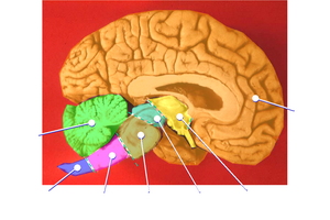
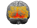
Human brain: sagittal section
|
![]() Also known as Human cerebrum
Also known as Human cerebrum
Subcategories
This category has the following 23 subcategories, out of 23 total.
B
C
F
- Fornix of the brain (28 F)
G
H
- Human striatum (46 F)
L
- Longitudinal fissure (30 F)
M
- Medial lemniscus (10 F)
O
P
S
T
Media in category "Human telencephalon"
The following 54 files are in this category, out of 54 total.
-
 1543,Vesalius'OlfactoryBulbs.jpg 290 × 308; 24 KB
-
 1543,Visalius'OpticChiasma.jpg 287 × 308; 24 KB
-
 201405 cerebrum.png 400 × 400; 29 KB
-
 Alina-grubnyak-1362365-unsplash.jpg 2,235 × 1,514; 442 KB
-
 Areabroca.jpg 512 × 366; 92 KB
-
 Blausen 0115 BrainStructures.png 1,600 × 1,429; 1.68 MB
-
 Brain human coronal section tags.png 628 × 413; 38 KB
-
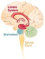
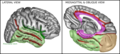
 Brain limbicsystem-es.svg 227 × 305; 43 KB
-
 Brain limbicsystem.jpg 200 × 263; 11 KB
-
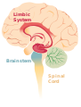 Brain limbicsystem.svg 240 × 316; 41 KB
-
 Brain visualization.jpg 500 × 500; 40 KB
-
 Brain-1.jpg 484 × 356; 58 KB
-
 Brodmann areas 17 18 19.png 256 × 192; 40 KB
-
 Cerebral Cortex location - he.jpg 283 × 337; 12 KB
-
 Cerebral Cortex location - pt.jpg 283 × 337; 46 KB
-
 Cerebral Cortex location.jpg 283 × 337; 17 KB
-
 Cerebral vascular territories midline.jpg 1,024 × 999; 127 KB
-
 Cingulate sulcus.png 170 × 140; 33 KB
-
 Constudoverbrain - 2 vie.png 539 × 698; 30 KB
-
 Constudoverbrain - 2.png 539 × 698; 11 KB
-
 Corteza Prefrontal.png 873 × 508; 17 KB
-
 Falxcerebri.jpg 650 × 636; 189 KB
-
 Gray and White matter of the cerebrum.png 1,728 × 2,304; 885 KB
-
 Gray657.png 550 × 358; 69 KB
-
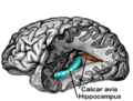 Gray739-emphasizing-calcar-avis.png 1,000 × 758; 997 KB
-
 Homunculus sensory.png 300 × 190; 36 KB
-
 Homunculus-ja.png 1,200 × 635; 197 KB
-
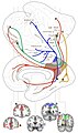
 Kea0003-papezkreis.PNG 584 × 254; 21 KB
-
 Lateral geniculate nucleus.png 300 × 259; 38 KB
-
 Limba sistemo sentekste.jpg 400 × 526; 56 KB
-
 Limba sistemo.jpg 400 × 526; 67 KB
-
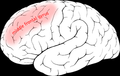 Middle frontal gyrus.png 300 × 190; 42 KB
-
 MRI of orbitofrontal cortex.jpg 705 × 672; 73 KB
-
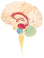
 Neocortex-thalamus-brainstem.jpg 420 × 291; 98 KB
-
 Otic ganglion 2.jpg 960 × 720; 92 KB
-
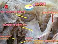 Otic ganglion.jpg 960 × 720; 87 KB
-
 Plaats hersenschors.png 283 × 337; 92 KB
-
_-_vista_inferiore.png/120px-Poles_of_cerebral_hemispheres_(it)_-_vista_inferiore.png) Poles of cerebral hemispheres (it) - vista inferiore.png 755 × 630; 311 KB
Poles of cerebral hemispheres (it) - vista inferiore.png 755 × 630; 311 KB
-
 Precentral sulcus.png 300 × 190; 27 KB
-
 Precuneus connectivity new.gif 343 × 447; 30 KB
-
 Precuneus connectivity.jpg 4,111 × 6,939; 81.65 MB
-
 PSM V42 D779 View of a Lobe of the Cerebrum.jpg 1,134 × 840; 99 KB
-
 Sistema BOS.jpg 426 × 202; 20 KB
-
 Some brain areas.png 989 × 412; 146 KB
-
 Superieur gyrus.png 554 × 516; 115 KB
-
 Superior frontal gyrus.png 300 × 190; 32 KB
-
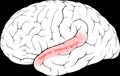 Superior temporal gyrus.png 300 × 190; 35 KB
-
 Surfacegyri.JPG 1,664 × 1,496; 143 KB
-
 Telencephalon2.png 800 × 455; 222 KB
-
 TempCapts.png 1,899 × 856; 880 KB
-
 ThalamicNuclei.png 960 × 720; 18 KB
-
 The Limbic System and Nearby Structures - John Taylor.jpg 800 × 755; 288 KB
-
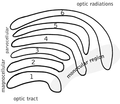
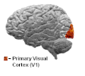 Visualcortex.gif 201 × 179; 10 KB
-
 Voorste centrale groeve.png 300 × 190; 31 KB


_-_vista_inferiore.png)


